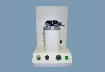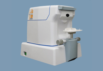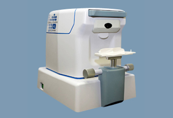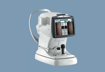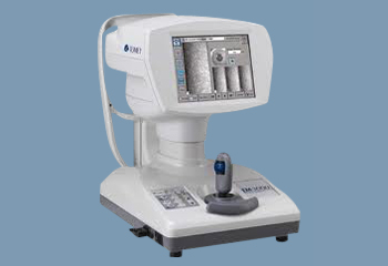
Specular microscopes are used specifically to diagnose corneal endotheliopathy. The condition can lead to visual impairment and is often linked to other serious problems or eye diseases like glaucoma and Fuchs endothelial dystrophy. Thus, specular microscopy is important to accurately diagnose the issue and determine the right course of treatment.
How specular microscopes work
Specular microscopes are noninvasive – they use computer-aided morphometry to assess the size, shape, and population of endothelial cells. The microscope projects light onto the cornea. Then, an image of the cornea is reflected and captured. The specular microscope analyzes the resulting image and displays a specular photomicrograph.
Benefits of specular microscopes
The greatest benefit of specular microscopes is their accuracy. They’re believed to be the best possible way to capture corneal endothelium photos and, in turn, enable doctors to provide a high standard of care. The findings from the specular photomicrograph can inform not just the patient’s primary eye doctor but also any other surgeons or optometrists brought in to assist with treatment.
Specular microscopes can be used before and after surgery to assess corneal health and look for any signs of trauma. And, in addition to their primary focus on corneal endotheliopathy, they can be used on corneal transplant patients and even patients with cataracts.
In some cases, a specular microscope can help ophthalmologists determine the right contact lens prescription for patients.
Additionally, for the patient, noninvasive procedures mean a more enjoyable exam experience.
In general, specular microscopes are an essential investment as the analysis they provide can help in a wide variety of cases.