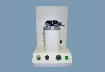
Konan’s compact CellChek EB Eye Bank Specular Microscope features a high resolution CCD camera that captures high quality images of the donor cornea. A built-in cell analysis system on the Konan CellChek EB provides rapid and accurate determinations of cell count, polymegethism (cell size variations) and pleomorphism (cell shape variations).
The Konan CellChek EB features XYZ and rocking platform mechanisms making tracking of endothelial cells easy. The built-in pachymeter allows measurement of corneal thickness, which is useful for evaluation of corneas that have undergone refractive surgery. The Konan CellChek EB can analyze the cornea either from a corneal chamber or directly from any vial, thus eliminating the risk of contamination and reducing costs. As an option, Konan provides a holder in which to place intact eyeballs for direct observation before tissue processing.
The built-in analysis system on the Konan CellChek EB has five different methods of performing cell counting, making it easy to analyze even corneas with harder to see cells. Donor information and cell analysis data with histograms are immediately displayed on the monitor; the endothelial image and all the necessary data can be printed out with the video printer.