Optical coherence tomography, also known as OCT, is a non-invasive imaging test and powerful diagnostic tool. It’s often referred to as an MRI for the retina. By using OCT, ophthalmologists can view each distinct layer of a patient’s retina and also map and measure each layer’s thickness. This precise retinal information can help diagnose several common eye conditions, such as central serious retinopathy and macular pucker, and it can inform treatment plans for retina diseases, like diabetic eye disease and age-related macular degeneration, and glaucoma.
Additionally, OCT can be used to further understand optic nerve disorders, as the OCT technology illuminates any changes to the optic nerve’s fibers. And OCT results can be compared to results from other testing to paint a fuller picture of a patient’s eye conditions.
How optical coherence tomography works
OCT takes high-resolution cross-section photos of the retina using low-coherence light waves, similar to the ways in which ultrasounds use sound to detect and diagnose conditions. Patients rest their chin on the OCT machine, with nothing touching the eye, and look at a light target. The physician calibrates the machine and the scan is complete in under 5 minutes.
Benefits of optical coherence tomography
OCT makes an excellent addition to any eye care office for many reasons:
- No radiation or X-rays are required, which means a safer standard of care for patients.
- OCT scans aren’t painful or uncomfortable, ensuring a positive patient experience.
- OCT scans have no known health risks.
- No pupil dilation is required.
OCT is a fast, effective, and patient-friendly way to diagnose and treat retinal diseases and conditions and maintain a high standard of patient care.
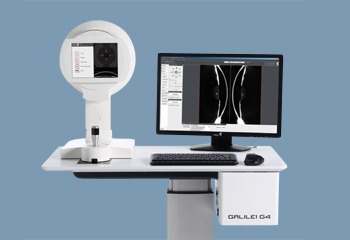
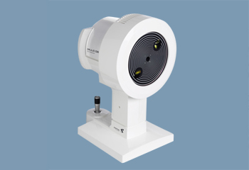
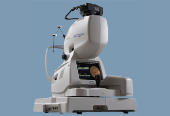
Heidelberg
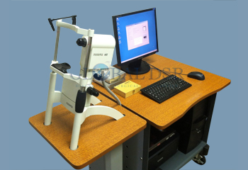
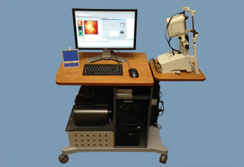
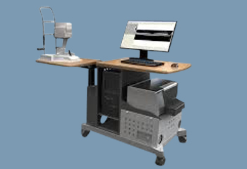
Optovue
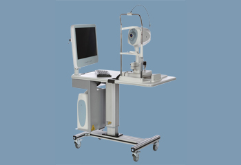
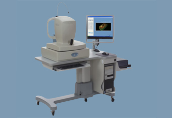
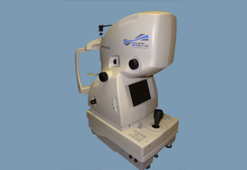
Topcon
Zeiss
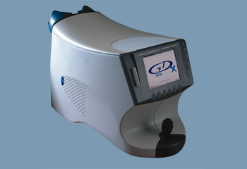
Zeiss GDx VCC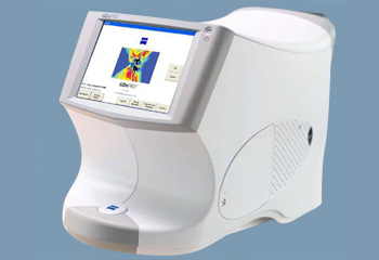
Zeiss GDx PRO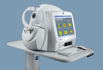
Zeiss Visante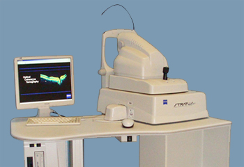
Zeiss Stratus OCT-III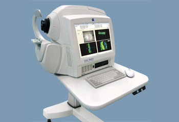
Zeiss Cirrus 4000
Zeiss Cirrus 5000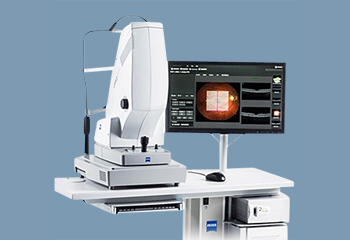
Zeiss Cirrus Photo