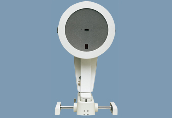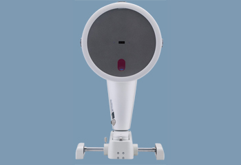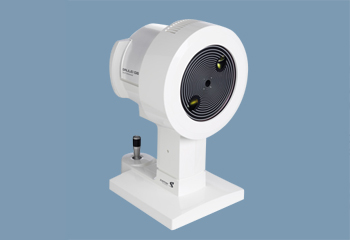
Schiempflug cameras employ a unique photography style to extract important diagnostic data from the eye.
A Scheimpflug camera sits perpendicular to a slit beam, which makes it easier to create images of the anterior segment of the eye. The captured images are used to create an optical section of the lens and the cornea.
These images are digitized and combined to create a 3D model of the eye’s anterior segment. This model becomes the reference point for all necessary information including:
Cornea condition (i.e. topographic thickness, curvature, etc.)
Height, angle, and volume of the anterior segment
This model is also used to generate pachymetry and topography for the cornea’s entire posterior and anterior surfaces. The model can even aid in quantifying cornea and lens opacity.
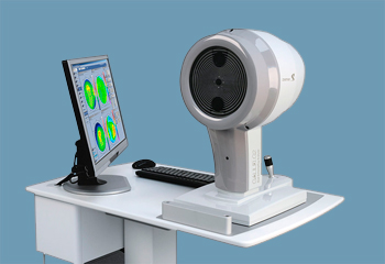
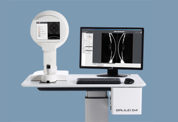
Scheimpflug cameras are valuable tools because they’re noninvasive, fast, and highly accurate. For example, the Pentacam, a rotating camera, takes just 2 seconds to generate a complete model of the anterior segment. In that 2 seconds, a 3D model may be calculated from up to 25,000 true elevation points.
Scheimpflug cameras are a vital resource for any modern ophthalmology practice. Physicians can gain an incredible amount of information from using these cameras, to do everything from treating cataracts and glaucoma to prepping for LASIK surgery to diagnosing serious eye conditions, like Fuchs endothelial dystrophy.
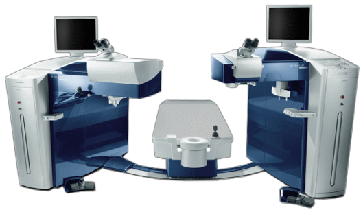
Below, you’ll find a curated selection of the industry’s top Scheimpflug cameras.