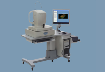
The Optovue RTVue OCT is an ultra-high speed, high resolution OCT retina scanner used for retina imaging and analysis. The Optovue RTVue OCT is based on the next generation Fourier-Domain Optical Coherence technology just emerging from clinical research in the last two years. The ultra-high speed and high resolution features enable the FD-OCT to visualize the retinal tissue with ultra-high clarity in a fraction of seconds.
Fourier-Domain OCT vs. Time-Domain OCT
The old generation OCT, is based on the Time-Domain OCT technology. In TD-OCT, there is a mechanical moving part, which performs the A-scan, and the information along the longitudinal direction is accumulated over the course of the longitudinal scan time. Due to the nature of the slow mechanical moving speed, the scan time in TD-OCT is very slow. For example, the current commercial retina scanner can only perform 400 A-scans/second. Because of the fast eye motion effect, it is not feasible to use TD-OCT to map the retina tissue. Therefore, it is limited in ocular applications which mostly require high repeatability and high data sampling rate, which must be completed in a fraction of a second.
In Fourier-Domain OCT, the technology used in the Optovue RTVue, the information in an entire A-scan is acquired by a CCD camera simultaneously. The A-scan acquisition rate is only limited by the CCD camera frame transfer rate and the computer calculation time to perform the Fourier transform of the CCD acquired raw data into A-scan information. Due to the fast CCD camera frame transfer rate and fast Fourier transform algorithm, FD-OCT, like Optovue RTVue, can perform 26,000 A-scan/second. This is a 65 times speed advancement over current technology.
Features:
• RTVue ultra-high speed: 65 time faster (26,000A-scan/s) for 3D and high density mapping
• RTVue two scan depth range: 2mm for imaging the macular and 2.3mm for imaging a tilt disk.
• RTVue two image modes:
o Vitreo mode: for better vitreoretinal tissue image
o Choriod mode: for better chorioretinal tissue image
• RTVue high transverse scan resolution:15µm (1/e2 spot size on retina)
• RTVue video fundus image: Fundus camera optical design for high quality NIR retina image
• RTVue Non Mydriatic imaging: The pupil size required for the fundus image is 3.0mm
• RTVue long working distance: 22mm from cornea to ocular lens